For decades, one of the deadliest parasites on Earth kept a secret. Once it infects human cells, it creates a protective layer that separates and protects it from the defense mechanisms of the host cell. This protective membrane, known as the parasitophorous vacuole, is essential for the ability of the pathogen to establish infection on humans. However, how this membrane divides simultaneously with the parasite remains a mystery. In 2025, a University of Florida Ph.D. candidate from the Department of Microbiology and Cell Science helped answer that question, and in doing so, identified a weakness that could one day become the target of a new treatment.

Javier Rosero studies Leishmania donovani, the parasite responsible for the deadliest form of leishmaniasis. Working alongside faculty mentor Dr. Peter Kima in one of the department’s state-of-the-art research laboratories, he has spent his doctoral career investigating how the parasite invades human cells, hijacks their machinery and proliferates. This year, he and Dr. Kima uncovered a critical piece of that process: a molecular mechanism the parasite cannot proliferate without.
For Javier, scientific discovery is driven as much by curiosity as experimentation. Reflecting on what motivates his research, he said:
A Disease the World Overlooks
L. donovani causes visceral leishmaniasis, the deadliest form of a disease the World Health Organization classifies as neglected, meaning it disproportionately affects poor and vulnerable communities and receives a fraction of the research investment given to higher-profile illnesses. Left untreated, it is fatal in the vast majority of cases. Leishmaniasis spreads through the bite of a sand fly and shows up across tropical regions of Asia, Africa and Latin America, including Colombia, which recorded more than 350 cases of the visceral form between 2017 and 2023, two of them children. It has also been identified in Texas, now making it a domestic concern as well as a global one.
In India, people call it kala-azar. Black fever.
Here is the problem the parasite has to solve to survive: blood is hostile territory, crowded with molecules, including antibodies, built to destroy it. So immediately after a bite, the parasite ducks inside a human immune cell called a macrophage and uses it as a hideout. Once inside, it wraps itself in a protective membrane called a parasitophorous vacuole. To reproduce, that membrane has to divide right along with the parasite. For decades, no one knew how.
“To observe the world at a microscopic scale is to reveal secrets of larger mysteries. I work with cells called macrophages, which are a subgroup of white blood cells. These cells are tasked with engulfing potential threats to your overall health. They were first discovered by Élie Metchnikoff, a zoologist in training who first observed through a microscope these cells engulfing other smaller cells. He wrote in his diaries that this observation was so exciting to him, that he could not sleep for days. It has been 144 years since he attested this, and I profoundly resonate with his experience, as I have had similar sleepless nights. Such is the scientific endeavor.”
The Discovery
Working alongside Dr. Kima, who has studied leishmaniasis for more than two decades, Javier helped identify the mechanism behind that division. The pair discovered that L. donovani recruits the host cell’s own endosomal sorting complex required for transport, known as ESCRT, a system the cell normally uses for membrane repair and its own division, and repurposes it to split the vacuole in two. One ESCRT-associated protein, ALIX, turned out to be especially critical to the process. Javier describes the complex, more simply, as the parasite’s molecular scissors: the tool it depends on to cut its way toward dividing.
When Javier’s team removed that machinery in experiments, the parasite got stuck. It could not divide, could not multiply inside the cell.
That is not a cure. It is a vulnerability, a specific piece of machinery a future drug could target to stop the parasite from reproducing at all. The findings, published as a first-author paper in PLOS Pathogens in September 2025, were the first to implicate ESCRT machinery in the division of an intracellular vacuole of any kind, a discovery with implications reaching beyond leishmaniasis into how other pathogens, from bacteria to viruses, may hijack the same cellular toolkit to survive inside human cells.
As Javier puts it, science does not move in shortcuts. This is one fundamental piece of a much larger puzzle, and the next step is translating the finding into an actual therapeutic strategy, including early work evaluating a class of chemical compounds, some already used to treat inflammatory bowel disease and studied for their effects on HIV, that may block the parasite’s ability to load infectious cargo into the vesicles it uses to communicate with neighboring cells.
“There is a quote from an old superman movie that I like a lot, it reads ‘Some people can read the novel War and Peace and come away thinking it is merely about adventures. Others can read the ingredients on a chewing gum wrapper and unlock the secrets of the universe.’ It refers to how imagination and creativity are essential to drive scientific progress forward. The discovery my mentor and I made started with a simple observation on the microscope that culminated in an important research article.”
Where Discovery Comes Into Focus
Discoveries like this are only possible because Javier can see what ordinary microscopy cannot. He has become the department’s go-to expert on advanced confocal microscopy and now trains other researchers, from undergraduates to postdoctoral fellows, on the instrument.
Among the department’s advanced research instrumentation is a Leica Stellar DMi8 confocal microscope, equipped with an Oko Lab enclosure that maintains precise temperature control and a Bold Line gas induction system that regulates carbon dioxide and oxygen levels inside the chamber. That setup allows Javier to keep living cells alive and infected under the lens for hours at a time, including under low-oxygen, or hypoxic, conditions that mimic the environment inside an infected host. Paired with five objective lenses ranging from 5x to 63x magnification and Leica’s Lightning deconvolution software, the system produces sharper, more detailed optical sections than standard confocal imaging allows. The microscope is also equipped for fluorescence recovery after photobleaching and fluorescence resonance energy transfer, or FRET, techniques that let researchers track how proteins move and interact in real time inside a living cell.
In practice, that means Javier can watch the parasite invade a macrophage, hijack its machinery and divide, live, in three dimensions, rather than reconstructing the story after the fact from static images. It is the kind of research-grade instrumentation few doctoral programs in the country can offer, and it has made Javier something of an in-house resource across multiple labs in the department.


From Bogotá to the Bench
Javier was born in Bogotá, Colombia, in 1995. He arrived in Gainesville in 2019 to study biology, without any particular plan to end up chasing parasites. That came later, from a conversation with a friend in a microbiology Ph.D. program and an undergraduate class on parasitic disease that hooked him.
There is a more personal thread, too. In 2018, when he was 22, his younger sister, then 14, was diagnosed with cancer. It was, in his words, a hard experience that forced him to grow up fast and pointed him toward medicine and disease research. His path from that moment to a UF lab was significant enough that El País, one of Colombia’s leading newspapers, profiled it earlier this year.
He emailed Dr. Kima in the middle of the COVID-19 pandemic, asking to join his lab. Pandemic restrictions delayed things, but by April 2021 he was in, learning to culture the parasite and run graduate-level techniques as an undergraduate on a University Scholars scholarship. He trained rotating graduate students, an international postdoctoral fellow and a Ph.D. candidate in the methods he had picked up along the way. He graduated magna cum laude with honors in 2022 and was accepted the same year into the Ph.D. program in Microbiology and Cell Science. He completed a Master of Science along the way, earlier last year, on a Grinter Fellowship.
“My interest in research, in my opinion, started when as a kid I liked collecting bugs with my dad. He used to bring me beetles and spiders as well as other insects. I always wondered what allowed them to move and live in their own peculiar way. As a matter of fact, I always dreamt of finding a praying mantis in the wild. I recall thinking that their odd movements seen on TV were worth seeing live.”
Recognized by His Field, Quietly
Ask anyone who has worked with him and the same word comes up before any list of accomplishments: generous. Colleagues describe someone who shares his expertise freely, treats collaborative projects as shared wins rather than personal credit, and mentors without being asked twice. He has formally trained more than 50 members of his department, from undergraduates to incoming and current Ph.D. students to international postdoctoral fellows, and has been nominated for the department’s Best Graduate Teaching Assistant award.
He is not, by nature, someone who leads with his résumé. The résumé, though, speaks for itself.
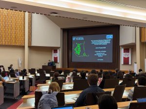
Beyond his PLOS Pathogens paper, Javier is a co-author on a 2024 review in GEN Biotechnology on engineered lab models for neglected tropical diseases and a 2026 study in Gut Microbes on how bacterial extracellular vesicles help control viral infection. His work was selected at the EMBO Workshop on ESCRT Biology in Switzerland in 2025 for an oral presentation, a significant distinction in a field where oral slots are reserved for the most compelling work. He has since presented at the International Society for Extracellular Vesicles conference in Puerto Rico and the 35th Molecular Parasitology and Vector Biology Symposium, and took second place in graduate student presentation categories at both the American Society for Microbiology’s Florida Branch meeting and UF’s Emerging Pathogens Institute Research Day, twice.


Mentorship Built to Match the Ambition
None of this happens in isolation. Javier credits the culture of the department, and specifically the mentors who pushed him to ask bigger questions before he had proof they would pay off.
His faculty mentor, Dr. Peter Kima, Ph.D., associate professor, has watched that curiosity define Javier’s approach to research.
“Javier has an inclination for exploring concepts and approaches in great depth. I enjoy watching him delve into the scientific literature to demystify the concepts we are exploring. This has allowed him to develop a detailed understanding of exceptionally complex processes. His understanding of imaging and confocal microscopy best illustrates that strength. Like an athlete of whom it is said that the game slows down as they become more proficient, scientists also develop a deeper understanding of their biological systems through careful study of the literature. Javier is a great role model for incoming students who have limitless potential but must accept the challenge to apply themselves.”
The same drive has also shaped Javier’s work as a graduate teaching assistant. Monika Oli, Ph.D., master lecturer, says his enthusiasm for microbiology extends well beyond his own research.
“Besides spending day and night in the research lab, Javier has been a graduate teaching assistant for many years. His passion for teaching parallels his passion for parasites. He goes above and beyond in the classroom, igniting a passion for parasitology and the unseen microscopic world in his students. This summer, Javier surprised me even more when he volunteered to conduct a special experiment for students in the new Clinical Microbiology Bootcamp. He infected macrophages with Leishmania, allowing each student to safely stain and observe their own parasitic vacuoles. I look forward to seeing where Javier’s career will take him.”
Discovery Starts at UF
Javier’s path is a working illustration of what a Ph.D. at the University of Florida is built to offer: mentorship from faculty who are recognized leaders in their field, access to instrumentation most doctoral programs cannot match and the room to carry a question all the way from a hunch to a named molecular target published in a leading journal.
Asked what he would tell a young person considering a career in science, Javier does not hesitate. It is a hard road, he says, but a deeply rewarding one, and it is more reachable than people think. He got here on scholarships. The opportunities are out there for anyone with the curiosity and the discipline to chase them.
“Choosing the Microbiology and Cell science department at UF has been the best choice for me to carry out my Ph.D. work. Being part of this department has helped my career tremendously. It has allowed for my creative freedom to materialize in ways I could not have ever even thought of. ”
From advanced live-cell imaging to hands-on mentorship, students in UF’s Department of Microbiology and Cell Science get the chance to contribute to discoveries that reach well beyond the lab bench, including, potentially, toward future treatments for diseases the rest of the world has largely overlooked.
Explore the On-Campus Ph.D. Program →
See more research from the department →
Interested in graduate school? Request more information →
 3
3