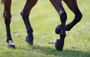
 Like the cornerstone of a building or the wheels of an automobile, the feet and legs serve as the foundation of the horse. Uniquely designed to support weight, resist wear, absorb tremendous shock, provide traction and assist in pumping blood, the horse’s foot and lower leg is without a doubt one of the greatest engineering feats in nature.
For horses to perform to their fullest potential, the feet and legs must be healthy and maintained in an impeccably sound condition. To help prevent disease, avoid unnecessary injury, keep horses sound and healthy, and allow horses to perform to their optimum ability, it is important for breeders, owners, and trainers to have a thorough understanding of the structures and functions of the equine foot and lower leg.
Approximately two-thirds of the horses’ weight is supported by the front legs. Bone is constantly rebuilding itself in response to conformation, training and nutrition and can either grow in a natural and desirable manner or an unnatural undesirable manner. When a column of bone experiences unequal stresses, the result is unequal growth. Regular trimming and shoeing by a knowledgeable farrier can do much to ensure uniform weight bearing of the leg bones.
Trauma to the leg and hoof bones may cause an increase in circulation and lead to an abnormal bony growth known as an osteophyte. When other factors restrict blood flow and circulation is decreased, degeneration of bone (known as rarefaction) may be the result. When the periosteum (sheath which covers the leg bones) is compromised, another abnormal bony growth known as periostitis results.
Cannon bone
 The cannon bone functions as a lever, and plays a direct role in determining the speed of a horse. The flat upper end of this oval shaped bone forms a large working surface for the knee bones. Designed to partially support the weight of the horse’s leg and withstand the powerful forces of work, the cannon bone is remarkably strong and not easily injured. There is no man-made structure of similar proportions that can withstand the force placed on it at speed. A relatively short cannon bone in relation to a long pastern allows the creation of leverage for speed and reduces concussion to the upper leg. However, shin splints can occur if the front of the cannon bone becomes irritated.
Splint bones
These two icicle shaped bones are located on either side of the rear of the cannon bone. In addition to providing a cradle in which the knee rests, these bones protect ligaments, tendons, blood vessels, and nerves. They also add to the structural integrity of the leg. “Splints” hard bumps formed by calcification between the splint and cannon bones are more often located on the inside of the leg because the inside splint bones bear more weight of the horse’s knee. Once splints quit growing and become “set”, they are usually considered blemishes.
Sesamoid bone
Located next to the cannon bone at the rear of the fetlock joint, ligaments attach these small pyramid-shaped bones to the long pastern bone. The sesamoid bones perform three primary functions: (1) serve as a bearing surface for the flexor tendon, (2) strengthen the fetlock joint by adding support to the cannon bone, and (3) provide leverage to tendons and ligaments. An inflammation of these bones is known as sesamoiditis.
Long pastern bone/First phalanx (P1)
The long pastern bone is approximately one-third the length of the cannon bone and similar in shape. Located between the fetlock and pastern joints, the upper end of this bone has three groves which connect with the bottom of the cannon bone to form the fetlock joint. The function of the long pastern bone is to increase flexibility of the fetlock joint. Ideally, the joint formed between the cannon bone and long pastern allows no lateral movement, where the slightest variation can adversely affect the horse’s stride. Flexibility, length, and angle of the long pastern directly influence smoothness of gait. Horses with excessively long and sloping pasterns run a greater risk of developing bowed tendons. Hyperextension of the fetlock joint can produce trauma which may result in a bony growth at the upper end of the long pastern known as an osselet.
Short pastern bone/Second phalanx (P2)
This cube shaped bone is concave on the top with two depressions that cradle the long pastern bone. The lower end of this bone is convex with one depression and articulates with the coffin bone. The short pastern bone permits the foot to move side to side and twist back and forth allowing the foot to rest evenly on the ground. Bony growth (exostosis) located around the pastern joint is called “high ringbone”. When the abnormal growth is located at the coffin joint it is referred to as “low ringbone”. “Interarticular” ringbone is between joints and “periarticular” ringbone is around joints.
Navicular bone/Distal sesamoid
Located on the rear surfaces of both the short pastern and coffin bone, the navicular bone plays a key role in the anti-concussive (shock absorbing) mechanism along with its ligamentous attachments. The deep flexor tendon passes directly underneath, and the navicular bone serves as a leverage point for the tendon so that it may easily slide over the bone.Problems created in this area either by concussion or compression are referred to as “navicular disease”.
Coffin bone/Third phalanx (P3)
This bone is the farthest out from the body and is completely enclosed in the hoof. Interaction between the coffin bone and the surrounding hoof structures serves as a shock absorber for the horse in motion. A major function of the coffin bone is to provide for the attachment and protection of blood vessels and nerves. Additionally, this bone also provides the point of attachment for the tendons that move the lower leg. The coffin bone is very porous and lightweight and can become irritated by unequal weight bearing, concussion, pressure or trauma that could result in abnormal bony growth called “pedal osteitis”.
Hoof
 The hoof is a cornified epidermis similar in makeup to the human fingernail and contains no nerves or blood vessels, but relies on corium (an inner layer) to provide the circulation and sensitivity necessary to maintain a healthy foot.
Corium
This internal component contains a massive supply of blood vessels and functions as the main nutritional source of the hoof. These blood vessels tied together with nerves form a sensitive layer which is attached to the inside of the hoof wall and coffin bone.
Bulbs of the heel
These structures are located at the back part of the ground surface of the foot, behind the angle of the hoof wall. They receive internal support from the digital plantar cushion.
Digital plantar cushion
This wedge shaped structure is composed of elastic tissues and some cartilage. The plantar cushion lies inside the lateral cartilage and is enclosed by the coffin bone, navicular bone, and the deep flexor tendon. As the name implies, this structure serves to lessen concussion to the foot. When this cushion is compressed by the pastern bones and frog it is forced outward and upward against the lateral cartilages with equal intensity.
The frog
 Often referred to as the horses’ second heart, the frog is a rubbery cushion, triangular in shape. It functions to aid in traction, circulation, and is an integral part of the horse’s shock absorbing mechanism. Many unhealthy, disease related conditions of the frog are the result of over trimming, dryness, uncleanliness and/or neglect.
The wall
The wall encompasses the entire foot and all its inner parts. Continually growing downward from the coronet band, the wall is divided into three general areas, the toe, quarter and heel. The wall is elastic in its makeup and is both strong and flexible. It bears most of the horse’s weight and is subject to wear, trauma and environmental conditions. The inside of the wall is made of “horny” laminae which are interwoven with “sensitive” laminae. This unique connection allows the hoof to grow downward while still maintaining its attachment to the coffin bone.
A thorough understanding of the preceding structures should help horsemen more accurately discern lameness problems of the hoof and/or leg. Happy feet make happy horses and happy horses make happy owners.
 1
1
